Information
Anatomy of the knee joint
The knee joint consists of three bones: the femur (FEMUR), the tibia (TIBIA) and the patella (PATELLA). The cap sits on the front of the knee, on top of the femur, and slides down into a "groove" when the joint is flexed. It strengthens the work of the quadriceps. The rubbing surfaces of the three bones are covered with articular cartilage - a tissue that ensures the smooth movement of the joint with a low coefficient of friction. The cartilage together with the menisci represent the "shock absorber" of the knee joint. Ligaments are strong and elastic collagen structures that connect bones and provide stability to the joint.
Four major and several other smaller ligaments connect the femur to the tibia:
- 1. Anterior cruciate ligament - Anterior cruciate ligament - located in the middle of the joint and mainly prevents the lower leg from moving forward and to the side relative to the thigh.
- 2. The posterior cruciate ligament (Posterior cruciate ligament) is located inside the joint and mainly prevents the lower leg from moving backwards relative to the thigh.
- 3. Fibular (external) lateral connection (Fibular collateral ligament) - located on the outside of the knee and protects the lower leg from displacement relative to the axis of the lower limb.
- 4. Tibial (internal) lateral ligament (Tibial collateral ligament) - this ligament is located on the inside of the knee and protects it from displacement relative to the axis of the lower limb.
The menisci are located between the two large bones in the knee joint. They are two - internal medial (Medial meniscus) and external lateral (Lateral meniscus). These are crescent shaped cartilages. The inner one is large and fused at its periphery with the articular capsule and tibia major, and the outer one is small and free, fused only with the tibia major. The menisci distribute 60% of the load in the knee joint in the various degrees of knee flexion, reducing the contact area of the cartilage surfaces. They also have an important function for the stability of the joint in all its movements. Cartilage is a fundamental structure in all joints, including the knee, and its condition plays a leading role in all joint functions.
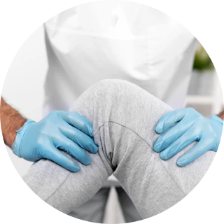
About knee arthroscopy
Knee arthroscopy is most often performed to diagnose and treat problems such as inflammation, injury, trauma, degenerative problems, and more. It can also be used to take a biopsy to clarify the cause of less common diseases (gout, rheumatoid diseases, tumor processes, etc.).
Arthroscopy is useful in the evaluation and treatment of the following conditions:
- Meniscus tear
- Injury to articular cartilage
- Removal of free bodies and cysts
- Anterior and posterior cruciate ligament (ACL/PCL) reconstruction
- Patellofemoral pain syndrome
- Inflammation of the knee joint
- Diagnosis and treatment of degenerative (age-related) changes in the knee
- General diagnostic objectives
Preparation for the procedure
1 week before surgery:
- It may be necessary to stop anticoagulants (aspirin, syntrom, etc.).
- Ask your doctor which medications you are allowed to take on the day of surgery.
- You should not drink large amounts of alcohol (more than 1-2 glasses per day).
- If you smoke, it is recommended that you stop, as smoking can delay healing.
- Tell your doctor about any cold, flu, fever, cold sores, or other illness you had just before surgery.
On the day of surgery:
- You will be asked not to eat or drink for 6-12 hours before the procedure.
- Take your prescribed medication with a small sip of water.
What does knee arthroscopy consist of?
Arthroscopy can last from 15 minutes to more than an hour, depending on the condition of the knee joint and what surgical procedures need to be performed.
There are four different types of anesthesia that can be used for knee arthroscopy:
- Spinal anesthesia - also called regional anesthesia. The pain-relieving drug is injected into a specific location around the roots of the spinal nerves. The patient remains awake but feels no pain from the waist down.
- General anesthesia - the patient is sedated through an intubation tube in the trachea and is unconscious during the procedure.
- PNB (peripheral nerve block) – the analgesic is injected along the course of the nerves that are responsible for the knee.
- Combined anesthesia - an analgesic is administered locally in the knee and the patient is put to sleep through a venous route.
Different methods of anesthesia have their advantages and disadvantages, which are discussed between the anesthesiologist and the patient. After the anesthetic takes effect, the surgeon makes several small punctures (4-5 mm) into the knee joint. A special liquid is poured into the joint and then the arthroscope (micro camera) is inserted.
Using the arthroscope and a monitor, the surgeon examines all intra-articular structures. Mini instruments are used to repair damaged tissue or remove material that interferes with movement or causes pain in the knee joint. Once complete, the surgeon aspirates the fluid from the joint and sutures the accesses. Then a bandage is made around the knee.
After the procedure:
Most patients go home on the day of surgery. You may need pain medication to deal with the discomfort of wearing off the anesthetic.
It is good to make arrangements in advance with a loved one to drive you home. It's good to have someone by your side during the first 24 hours.
You will be given some advice on how to care for the healing wounds. You will also be given a date for follow-up examinations (usually the 7th and 12th day).
The length of time it takes for self-dissolving stitches to disappear depends on what your condition was. However, with this procedure they usually disappear after about six weeks.
Recovery
Recovery from knee arthroscopy is much faster than traditional open surgery. However, it is important to follow your orthopedic surgeon's instructions carefully. It may be necessary to wear compression stockings that help maintain circulation, as well as various orthotics (knee braces, splints).
It is important to keep your knee clean and dry for about 10 days after surgery. In no case do not soak the knee in a bath until the wounds are completely healed. Do the exercises recommended by your physiotherapist as they will help improve the range of motion and strength in the knee.
There may be pain and swelling in the knee, most often one to three weeks after surgery. In some conditions (arthritis, degenerative changes, after micro fractures and larger and complex reconstructive operations) this may last longer. Try to keep your leg elevated above chest level on a chair or pillow when resting. A cold compress (ice wrapped in a towel) 3-4 times a day is recommended. This will help reduce swelling and bruising. It is advisable not to apply ice directly to the skin as it can damage it.
Follow your doctor's advice about driving. You should not drive until you are confident that you can make an emergency stop without discomfort. This usually happens about one to three weeks after surgery.
You should be able to resume your normal activities after six to eight weeks, depending on the severity of the problem and your fitness level (except for reconstructive surgeries of the ACL and SCL and some fractures).
Physiotherapy
Strengthening the thigh muscles is the most important. Swimming and cycling (exercise bike) are excellent ways to build these muscles and improve knee mobility.
Every patient is unique. The time it takes to recover depends on the type of injury, your fitness level and whether you had any complications.
After knee arthroscopy, small scars will remain on the knee from the incisions.
Possible complications
According to literature data, the total percentage of possible complications in arthroscopy is less than 0.5%. Most often, they are minor and transient (disappear completely in 5-7 days).
Complications
Potential postoperative problems include:
- Infection
- Formation of blood clots
- Accumulation of blood in the knee
Call your orthopedist immediately if you experience the following:
- A fever
- Shivering
- Persistent warmth or redness around the knee
- Constant or worsening pain
- Significant swelling in the knee Increasing pain in the calf muscles
Benefits of knee arthroscopy
Unlike open surgery, arthroscopy results in less pain and stiffness, fewer complications, a shorter hospital stay (if any), and a faster recovery.

 On this page, you can change your choices at any time after you have read and understood our