
Information
Introduction to CMV (Cytomegalovirus)
The incidence of CMV (Cytomegalovirus) infection in Bulgaria is unknown due to the lack of screening studies and behavioral protocols. CMV is the most common viral infection transmitted from a pregnant woman to her unborn child in the USA. It is also the most common congenital viral infection leading to consequences in the neonatal period and childhood. Approximately 40,000 newborns are born with congenital CMV infection in the USA each year, of which 6,000 have permanent consequences. Congenital CMV infection is responsible for approximately 25% of cases of newborns with hearing loss: 5 in every 1,000 fetuses born with CMV infection will die immediately after birth or in the first year of life; infected fetuses frequently develop epilepsy, intellectual disability, vision loss, disability, and various degrees of behavioral and cognitive deficit after birth.
Epidemiology
CMV is a DNA virus from the herpesvirus group.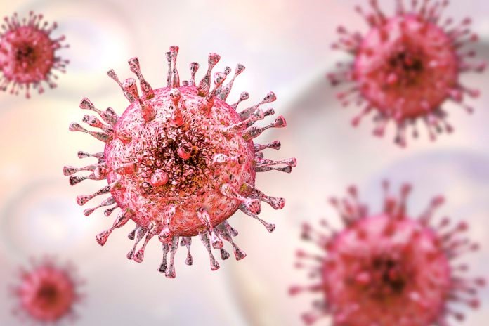 The initial development is an acute infection, after which the virus remains in a latent state in the body with subsequent relapses, typical of all infections in the herpesvirus group. Routes of transmission include: airborne droplets, from mother to fetus during pregnancy, and through sexual contact. It may be asymptomatic or present as a respiratory infection during pregnancy. The risk of fetal infection varies depending on whether an acute or recurrent infection develops during pregnancy.
Diagnostics
Due to the great significance of the infection, its early diagnosis during pregnancy is of great importance.
Diagnosis in the mother
Unlike other viral infections, testing for IgM and IgG antibodies is not sufficient. After the acute infection, IgM antibodies remain in circulation in the pregnant woman for a long time, sometimes almost a year. During a relapse, a surge in the secretion of class M antibodies in the patient's circulation is also observed. Differentiation of acute from chronic infection is achieved by testing the avidity of IgG antibodies. Avidity is the property of these antibodies to bind more strongly to the antigen over time. In acute infection, avidity values are low, while in chronic infection they are high.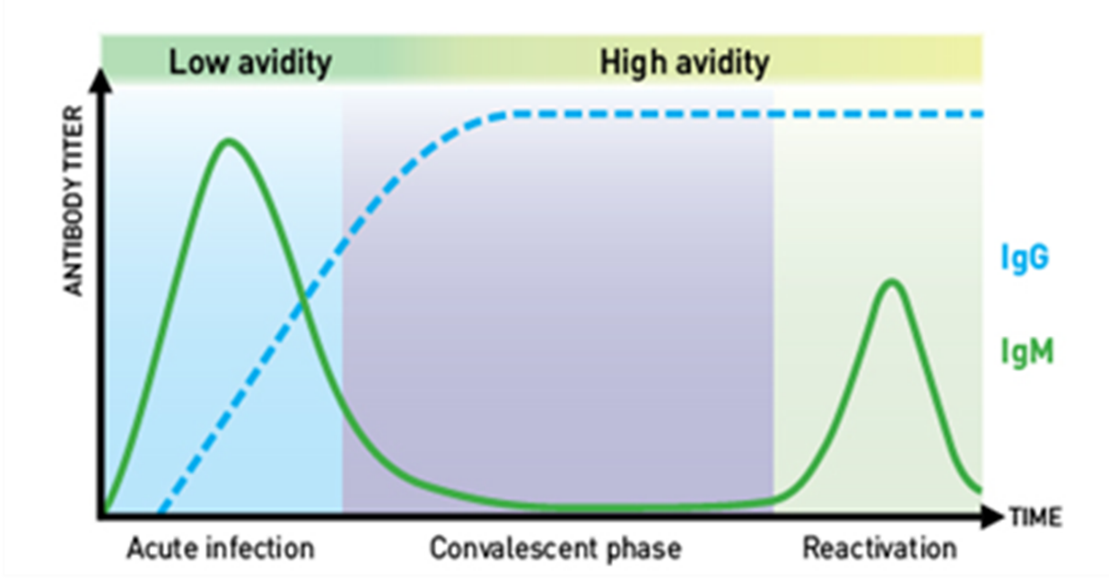
Changes in class M and G antibodies and IgG avidity during acute infection, chronic infection, and the convalescent phase.
Diagnosis in the fetus
In acute CMV infection, the fetus is infected in 40% of cases, and in recurrent infection in 0.5-2% of cases. The latest studies show that the greatest risk of intrauterine damage occurs when the infection occurs during the first trimester of pregnancy.
There is no completely accurate method for detecting fetal infection in utero. Sampling of amniotic fluid by amniocentesis and subsequent isolation of the virus by PCR can confirm the presence of fetal infection. Of the infected fetuses in patients with primary infection, 10-15% will have symptoms at birth, and 25% of newborns will develop symptoms by 2 years of age.
In patients with recurrent infection, the risks to the fetus are lower - approximately 1% of children will have symptoms at birth, and 8% will develop symptoms by 2 years of age.
Predicting which infected children will develop clinical symptoms after birth and will have permanent consequences is difficult. According to data from one of the major studies on the subject, after serial cordocenteses, thrombocytopenia below 114,000, the presence of β2-microglobulin, and high levels of CMV DNAemia - above 10,000 copies per ml in fetal blood - are considered poor prognostic markers. Ultrasound markers in monitoring CMV-infected fetuses also have prognostic significance.
In general, ultrasound markers are divided into two groups - CNS anomalies (ventriculomegaly, calcifications in various brain structures, interventricular septum, periventricular hyperechogenicity, periventricular cysts, hypoplasia of the corpus callosum, microencephaly, microcephaly) and non-CNS anomalies (polyhydramnios, oligohydramnios, IUGR, hyperechogenic bowel content, hepatosplenomegaly, cardiomegaly, hydrops).
In the presence of ultrasound anomalies outside the CNS, 4.4% of fetuses have clinical symptoms immediately after birth and in the early neonatal period, while in the presence of CNS ultrasound anomalies, 40.6% of fetuses have clinical symptoms immediately after birth and in the early neonatal period.
Clinical picture
In the mother, the infection most commonly presents asymptomatically or with symptoms of a respiratory infection.
In the fetus, hepatosplenomegaly, thrombocytopenia with petechiae, intracranial calcifications, low birth weight, microcephaly, hearing loss, and seizures are observed immediately after birth, either individually or in combination, expressed to varying degrees. In childhood, late consequences are observed - neurological symptoms of varying severity and visual impairments. CMV is the most common viral cause of intellectual disability and the leading non-genetic cause of sensorineural hearing loss.
Clinical case
A second-time mother was admitted on call with active labor.
Labor progressed rapidly with no evidence of fetal distress from the non-stress test performed. Notably, there was an abundant amount of green-stained amniotic fluid. The patient delivered a full-term fetus in critical condition: atonia, areflexia, no spontaneous respiratory effort, isolated muffled heart sounds, with livid discoloration of the skin on the head and marble-white skin on the body covered with petechiae. Chest X-ray revealed cardiomegaly and evidence of severe pneumonia. Despite full resuscitation measures, exitus letalis occurred at the 12th hour after birth. Of the extensive range of tests performed, the only positive results were IgM and IgG antibodies against CMV, detected in both the mother and the fetus.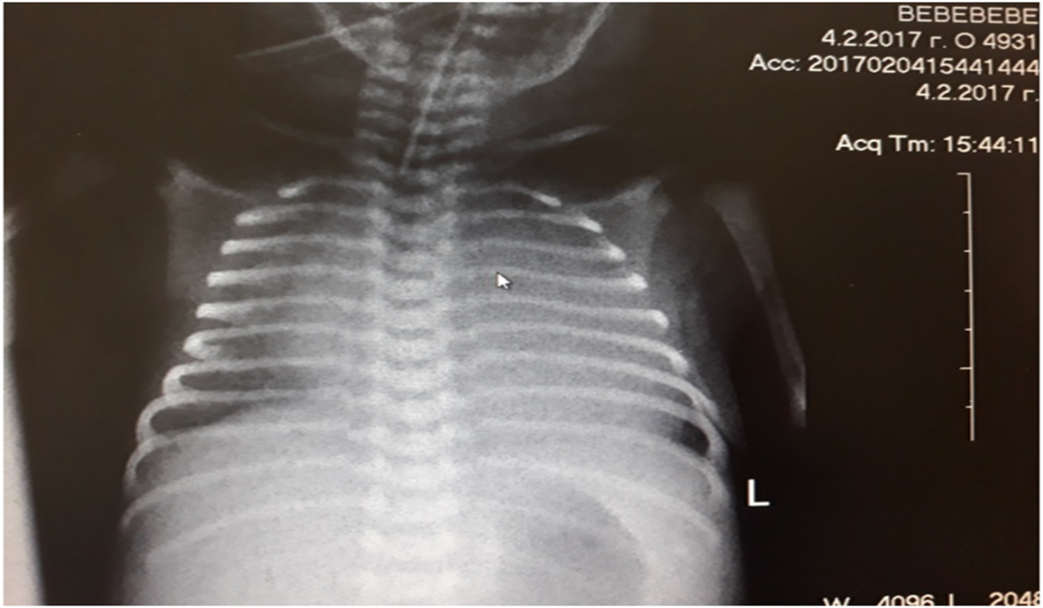 Cardiomegaly and severe pneumonia with an altered cardiothoracic index.
Within ten months of the first case, a total of four clinical cases were diagnosed, identified through mass screening among newborns:
- One ended in exitus letalis
- One - with bone marrow suppression - pancytopenia with Neu 0.0
- One - with cerebral calcifications and partial hearing loss
- One - with cerebral calcifications without symptoms in the early neonatal period
Conclusion
CMV infection is a socially significant disease due to its high incidence, the severity of clinical manifestations in affected fetuses, and the permanent disability of some children with intrauterine CMV infection. Knowledge of diagnostic methods, early identification of at-risk patients during pregnancy, and proper referral for consultation with specialists would help ensure adequate management of affected patients.
Bibliography
- Diagnosis and antenatal management of congenital cytomegalovirus infection. American Journal of Obstetrics and Gynecology. 214, 6 (2016), B5-B11.
- Fabbri E, Revello MG, Furione M, et al.: Prognostic markers of symptomatic congenital human cytomegalovirus infection in fetal blood. BJOG. 2011;118(4):448-56. 10.1111/j.1471-0528.2010.02822.
- Benoist G et al, 2008, British J Obstet Gynaecol, 115: 823-9
- Lanzieri TM, Leung J, Caviness AC, et al. Long-term outcomes of children with symptomatic congenital cytomegalovirus disease. J Perinatol. 2017;37(7):875-880.


 On this page, you can change your choices at any time after you have read and understood our